
by Tammy Stearns, MS, RDMS, RVT, RT(R), FSDMS, FAIUM, LAS
Techniques to Improve the Accuracy of Measuring in the First Trimester
Caliper placement can be a daunting task when measuring something smaller than a coffee bean. Sometimes, it appears that the calipers are bigger than the baby itself making it even more difficult. There is also the decision of if the caliper goes inside or outside with each miniscule change, making a difference in the data obtained.
There are a few techniques that can be utilized to help ensure that the measurements obtained are the most accurate possible. These same techniques improve resolution, while decreasing the uncertainty of the caliper placement and by providing more distinct borders.
When beginning the scan, be sure that the correct exam type is selected along with the best frequency for the client being scanned. Prior to placing the caliper, be sure that the images have the highest resolution possible by ensuring that the structure of interest is in the center of the screen. This can be achieved by applying pressure to either side of the transducer to move the image from right to left. The image can be centered vertically by either increasing or decreasing depth. Once the structure is imaged in the center of the screen, depending on the size of the structure, the ZOOM or RES can be utilized to enlarge the image of the structure. Also, be sure that the transmit or focal zone is level to the structure of interest or just below it to allow for the highest resolution.
Mean Sac Diameter
The Mean Sac Diameter (MSD) is one of the most important measurements in first-trimester obstetric ultrasound. It is used to estimate gestational age, assess early pregnancy development, and determine pregnancy viability when an embryo is not yet visible. The Mean Sac Diameter (MSD) is the average of the three internal dimensions of the gestational sac: Length (Longitudinal orientation-taken right to left), Height (Anterior-Posterior) and Width (Transverse orientation-taken right to left). The machine will add these measurements and divide them by 3. Measurements are made from the inner edge to the inner edge of the gestational sac (inside the echogenic trophoblastic ring). Be sure to include the fluid-filled cavity without including the trophoblastic ring, the decidual reaction, or the chorion.
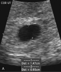 Longitudinal with AP and length measurements.
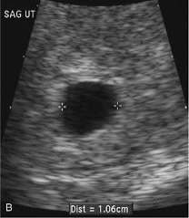 Transverse with width measurements.
- MSD ≥25 mm with no embryo = pregnancy failure.
- The yolk sac is usually visible when the MSD reaches 8–10 mm.
- CRL is the most accurate measurement once an embryo is present.
Crown-Rump Length
Once the embryo is visible, Crown Rump Length (CRL) should be used instead of the mean sac diameter for estimating gestational age. The CRL is the most accurate ultrasound measurement for determining gestational age during the first trimester. It measures the length of the baby from the top of the head (crown) to the bottom of the torso (rump), excluding the yolk sac, limbs, and umbilical cord. It is considered the most accurate measurement for pregnancy dating between 6 weeks and 13 weeks 6 days, being accurate to approximately ±3–5 days when measured correctly in the first trimester. Past this time, the baby tends to curl up on itself making it difficult to obtain an accurate linear measurement.
The baby should be imaged in a true midsagittal (long-axis) plane of the baby, not the mother. Measurements will be taken from the outer edge of the crown to the outer edge of the rump being sure not to include the yolk sac, limbs, and umbilical cord. The CRL is the preferred method for establishing the estimated due date (EDD) in the first trimester. Once an EDD is assigned from the first CRL, it is generally not changed based on later ultrasounds unless there is a compelling clinical reason.
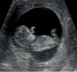
- CRL is used together with cardiac activity.
- CRL ≥7 mm with no cardiac activity is diagnostic of pregnancy failure.
- CRL <7 mm with no heartbeat is suspicious but not diagnostic; repeat ultrasound is recommended in 7–10 days.
Femur Length
After 13 weeks, the Femur Length (FL) can be utilized to give an estimate of gestational age. It should be noted that the scans performed in Pregnancy Help Medical Clinics are Limited Obstetrical Ultrasound scans. Therefore, femur length will give a general estimate of gestational age with the pregnancy to receive a referral for a follow-up complete obstetrical scan to include abdominal circumference, head circumference, and biparietal diameter if the mother decides to continue with the pregnancy. The FL is the measurement of the ossified diaphysis (shaft) of the fetal femur. The femoral head, greater trochanter, distal femoral epiphysis (if present), or cartilaginous ends of the bone are not included in the measurement.
The femur length is obtained in a view in which the femur is horizontal on the ultrasound screen and perpendicular to the ultrasound beam to minimize foreshortening. Be sure to visualize the entire diaphysis measuring from the outer edge of one ossified end to the outer edge of the other ossified end. The bone should appear as bright (echogenic) and as close to 90° to the ultrasound beam as possible to minimize foreshortening.
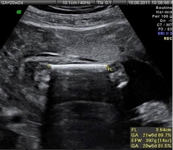 Femur Length- Only include the shaft of the femur.
- Femur should be perpendicular to the sound beam.
- Calipers should extend from the outer edge to the outer edge of the ossified ends.
Measurement accuracy can be improved by following a few specific techniques to increase image resolution and allowing calipers to be placed appropriately. Image quality along with correct structure and caliper placement play significant parts in accurate measurements. Part of the purpose of the Limited Obstetrical Scan is to provide the mother with the information that she needs to make an informed decision. Estimated fetal age is one of those pieces of information.
